
Introduction:
Adenocarcinoma is the most common type of malignant tumor in the stomach, accounting for approximately 95% of all stomach tumors. These adenocarcinomas are classified into two types: cardia cancer and non-cardia cancer, based on their anatomical location in the stomach. The remaining cases are comprised of lymphomas, carcinoid tumors, and gastrointestinal stromal tumors.(1) While the incidence of stomach cancer has significantly declined over the past fifty years, it still presents a major medical challenge, particularly in the age group of 60 to 80 years, though it may also appear in younger individuals.(2)
In this clinical case, we discuss the diagnosis and treatment of a 50-year-old male patient diagnosed with signet-ring cell carcinoma of the stomach. The patient presented with epigastric pain, significant weight loss, and general fatigue. After clinical examination and a series of medical investigations, the patient was treated initially with chemotherapy, overseen by an oncologist, followed by complete surgical removal of the stomach (gastrectomy). Post-surgery, the patient was discharged from the hospital in good overall condition.
This case highlights the importance of a multidisciplinary approach, combining chemotherapy and surgery, in the effective management of advanced gastric cancer.

Case Presentation:
A 48-year-old man presented with non-metastatic epigastric pain that began 9 months ago in intermittent episodes. The pain worsened when the patient was hungry and subsided after eating. His symptoms were accompanied by poor appetite and vomiting after consuming fatty foods, without any fever or changes in bowel habits. The patient also reported a weight loss of approximately 10 kg over recent months, and there were no signs of gastrointestinal bleeding, such as melena or hematemesis. He had no significant medical history.
Clinical examination:
The clinical examination revealed the patient in good general condition with stable vital signs. The chest examination was clear and symmetrical, while the abdomen was soft and moved normally with respiration, showing no signs of tension or guarding.
Several differential diagnoses were considered, including gastric ulcer, stomach cancer, and gastritis. Initial diagnostic tests returned with normal results, leading the medical team to pursue further testing for a definitive diagnosis.
Diagnostic procedures:
After the patient’s visit to the gastroenterology clinic, a tailored pharmacological regimen was prescribed to treat a suspected gastric ulcer. However, the condition did not improve, and additional symptoms developed, including diarrhea and food avoidance due to fear of vomiting or pain. Consequently, an upper gastrointestinal endoscopy was deemed necessary.
A comprehensive series of laboratory investigations were performed, including blood glucose analysis, liver enzyme assays, viral hepatitis screening, renal function tests, and coagulation and bleeding profiles. All results were reported to be within normal physiological limits.
The upper gastrointestinal endoscopy identified a large, ulcerated mass along the lesser curvature of the stomach, beginning just below the cardia and extending to occupy most of the lesser curvature. Multiple biopsies were taken for histopathological examination. These findings confirmed the presence of a gastric mass, prompting further pathological evaluation.
Furthermore, the endoscopic examination revealed that the esophagus and pyloric opening appeared normal, while the ulcerated mass on the posterior gastric wall extended towards the pyloric orifice, obstructing a significant portion of the stomach cavity. A biopsy measuring 3 × 3 × 2 mm was collected, and the final diagnosis, based on histopathological analysis, was signet-ring cell carcinoma originating from the body of the stomach.
Initial measure:
After the patient was diagnosed with a gastric lesion, a CT scan with contrast injection was performed. The imaging revealed the presence of irregular medium-intensity thickening in the midsection of the stomach and in the curvature areas, with marked enhancement of the contrast material. Based on these findings, the patient was referred to the oncology clinic, where they adminstrated 4 cycles of chemotherapy.
Following the chemotherapy treatment, it was decided to repeat both the gastrointestinal endoscopy and CT scan.
The second upper gastrointestinal endoscopy identified a hoof-shaped ulcer with slightly raised, fragile edges, located on the posterior wall and the lesser curvature of the stomach body, approximately 2 cm from the gastric junction. The ulcer measured 2-3 cm in diameter and 4 cm in length, with notable inflammation and congestion of the mucosal lining in the stomach body.
The second CT scan showed that the stomach and intestines in the studied sections were within normal limits, with no pathological changes or abnormalities detected. Similarly, the CT scans of the brain, neck, chest, and abdomen showed that all organs were normal in shape, size, and density, and no abnormal masses, cysts, or nodules were identified.
These findings suggest that all examined organs were within normal limits, and there was no evidence of significant disease or health concerns in the brain, neck, chest, or abdomen.
Surgical intervention:
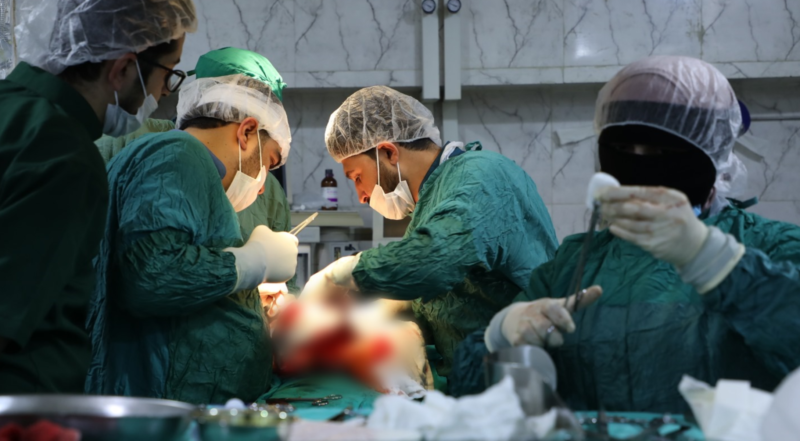
After initial examinations and chemotherapy, the patient was treated with a surgical intervention that involved a total gastrectomy. During the preoperative phase, an investigative laparoscopy was performed to assess the possible presence of peritoneal metastases. During this procedure, a biopsy was taken from suspected peritoneal implants, and a sample of peritoneal fluid (milky in appearance) was collected for pathological analysis.
Treatment Response and Surgical Outcomes:
The patient showed a good response to chemotherapy, allowing the medical team to proceed with the surgical plan. The entire stomach was successfully removed, and the excised tissue was sent for histopathological examination, which confirmed that the surgical margins were free of cancer (SRCC – free surgical margins). The peritoneal biopsy revealed a diagnosis consistent with non-specific peritonitis, and the analysis of the peritoneal fluid indicated it was benign inflammatory reactive fluid. However, metastatic involvement was identified in 4 out of 12 lymph nodes, classifying the patient’s condition as Stage IIIA.

Postoperative Course of Care:
Following the surgery, the patient remained in intensive care for 4 days to closely monitor his condition and ensure stability. Once his condition improved, he was transferred to the hospital ward to continue his recovery. On the eighth day post-surgery, the patient was discharged in good health.
Results and Follow-up:
The patient is currently in good general condition and maintains excellent health post-surgery, with ongoing follow-up care.
Discussion on Surgery and Chemotherapy in Stomach Cancer:
Adenocarcinoma, a significant subtype of stomach cancer, accounts for approximately 95% of all gastric cancer cases. It arises from the glandular cells of the stomach lining and is primarily divided into two types based on its anatomical location: cardia adenocarcinoma (situated near the esophagus) and non-cardia adenocarcinoma (found in the lower part of the stomach). This classification is critical for determining the treatment approach and prognosis of the disease .(1)
In 2020, around 1.1 million new cases of stomach cancer were diagnosed worldwide, making it the fifth most common cancer globally. Additionally, stomach cancer is the fourth leading cause of cancer-related deaths, responsible for an estimated 800,000 deaths annually. There is a significant geographic variation in the incidence of stomach cancer. East Asian countries, particularly China and Japan, report some of the highest rates of adenocarcinoma. In contrast, North America, and Northern Europe experience much lower incidence rates, highlighting the influence of environmental, dietary, and genetic factors on the disease.(3)
Risk Factors for Stomach Cancer:
The main risk factors contributing to the development of stomach cancer include:
- Infection with Helicobacter pylori (H. pylori), a known carcinogen in gastric adenocarcinoma.
- Dietary factors, particularly a diet high in salt, which is associated with an increased risk.
- Lifestyle factors, such as smoking and obesity, which elevate the likelihood of developing gastric cancer.
- Socioeconomic status and conditions like chronic gastritis, which have been linked to higher incidence rates in certain populations.(4)
The five-year survival rate for intestinal adenocarcinoma remains low, often below 20%, largely due to the late-stage diagnosis when symptoms become apparent. Recognizing these risk factors and understanding the dynamics of the disease are essential for developing targeted prevention strategies and improving methods for early detection of adenocarcinoma globally.
Chemotherapy in Stomach Cancer: Stomach cancer generally responds well to cytotoxic chemotherapy, and neoadjuvant chemotherapy—administered before surgery—has been shown to significantly improve surgical outcomes. Most patients are recommended to receive preoperative chemotherapy, and various regimens have been rigorously evaluated in clinical studies. This therapeutic approach offers several key theoretical advantages:
- Better integration of multimodal therapy, allowing for a coordinated approach to treatment.
- Reduction of tumor size prior to surgery, making the surgical procedure less complicated and more effective.
- Early treatment of micrometastatic disease, addressing cancer cells that may have spread undetected.
- Assessment of tumor response to chemotherapy before surgery, allowing clinicians to adjust the treatment plan based on the tumor’s reaction.
This strategy has proven beneficial in improving overall survival rates and enhancing the effectiveness of surgical interventions in treating stomach cancer.
Surgical intervention, particularly gastrectomy, remains a cornerstone in the treatment of localized stomach cancer. Total or partial gastrectomy can be performed depending on the tumor’s location and extent. However, surgery alone is often insufficient, especially in advanced stages of the disease. Combining surgery with chemotherapy—either in the neoadjuvant (preoperative) or adjuvant (postoperative) setting—has been shown to improve survival outcomes.
Chemotherapy plays a vital role in the management of stomach cancer by reducing the tumor size, enabling a more effective surgical resection, and targeting micrometastatic disease. For patients with locally advanced cancer, neoadjuvant chemotherapy is employed to downsize the tumor and facilitate complete resection.(5) Studies have demonstrated that this approach can increase the overall survival rate and reduce the likelihood of recurrence compared to surgery alone.
In advanced stages or cases with metastatic involvement, chemotherapy is the primary modality of treatment, aiming to control tumor growth and alleviate symptoms. Several chemotherapeutic regimens, such as 5-fluorouracil-based combinations, have proven effective in improving survival outcomes for patients with advanced gastric cancer.
Randomized clinical studies conducted in the UK demonstrated a significant improvement in survival rates among patients who underwent surgery in conjunction with perioperative chemotherapy, compared to those who underwent surgery alone. This improvement was especially pronounced in advanced tumor cases, where preoperative chemotherapy had a notably positive impact on patient outcomes.
Patients who received chemotherapy in conjunction with surgery exhibited a 13% increase in 5-year survival rates compared to those who had surgery alone, as well as a 25% reduction in the risk of death. These findings strongly support the integration of surgery and chemotherapy as an effective treatment strategy for stomach cancer.
The study specifically highlighted the efficacy of the ECF regimen—a combination of 5-fluorouracil, Epirubicin, and Cisplatin. This regimen, when administered in tandem with surgery, significantly reduced tumor size and improved overall survival for patients with resectable adenocarcinoma of the stomach or lower esophagus. Additionally, the study demonstrated a substantial improvement in survival rates and a decreased likelihood of disease progression, further validating the benefits of perioperative chemotherapy in managing gastric cancer.(5)
Benefits of Preoperative Chemotherapy:
- Tumor Size Reduction: Chemotherapy administered before surgery can effectively shrink the tumor, making initially non-resectable tumors excisable and improving the likelihood of achieving a complete resection (R0). This approach benefits patients with both clearly resectable disease and those with locally advanced tumors that may not be resectable at first.(6)
- Applicability to Various Patient Groups: Preoperative chemotherapy is applicable not only to patients with resectable tumors but also to those with non-excisable yet non-metastatic tumors, providing a broader scope for treatment.
- Early Targeting of Micrometastases: Administering chemotherapy before surgery allows for the early treatment of potential micrometastases, which is especially advantageous for patients at high risk of distant metastases. This systemic approach helps eliminate hidden metastatic cells that surgery alone may not address.
- Improved Tolerance and Completion Rates: Patients often tolerate chemotherapy better before surgery than after, as their nutritional status and overall performance are generally better before undergoing major surgical procedures. This can lead to higher completion rates of the full chemotherapy regimen, maximizing its effectiveness.
Preoperative chemotherapy, therefore, enhances the chances of successful surgical outcomes and systemic disease control, offering a comprehensive approach to managing stomach cancer. Evidence from multiple randomized trials supports this multimodal approach as superior to surgery alone for many patients with locally advanced stomach cancer.
Recommendations:
Laparoscopy and Peritoneal Lavage: Laparoscopy has emerged as a valuable tool in the staging and classification of stomach cancer, particularly in patients with large, locally advanced tumors. This procedure is essential for detecting small metastases in the peritoneum and liver that may not be visible on a CT scan. In addition, peritoneal lavage can provide critical information, as the detection of malignant cells in the peritoneal fluid is associated with a poor prognosis. In such cases, surgery is rarely beneficial, and guidelines suggest that “surgery should be postponed in patients with positive peritoneal lavage results but without visible peritoneal metastases.”(7)
Cancer Monitoring After Treatment:
Monitoring patients after treatment for adenocarcinoma of the stomach is crucial for detecting disease recurrence, managing complications, and ensuring patient well-being. Current guidelines for post-treatment follow-up include the following:(8)
- Clinical History and Physical Examination: These should be conducted every three to six months during the first two years and then every six to twelve months for the subsequent three to five years.
- Laboratory Tests: Regular complete blood counts and chemistry panels should be performed based on clinical indications to monitor overall health and detect any abnormalities.
- Nutritional Deficiencies: Monitoring for iron deficiency and vitamin B12 deficiency is particularly important in patients who have undergone procedures such as total gastrectomy. Nutritional supplementation should be provided as needed.
- Endoscopic Monitoring: For patients who have undergone partial or total gastrectomy, upper gastrointestinal endoscopy is recommended based on clinical indications to facilitate early detection of new lesions or complications.
- Imaging Studies: CT scans of the chest, abdomen, and pelvis with contrast should be performed every six to twelve months for the first two years and then annually for up to five years. This helps in the detection of recurrence or metastasis.
- Nutritional Support: Guidelines from the European Society of Medical Oncology (ESMO) emphasize the importance of nutritional follow-up post-treatment. This includes monitoring for vitamin and mineral deficiencies, especially due to the potential impact of surgical interventions on nutritional intake.(9)
- Additional Considerations: Routine evaluations should also address the potential need for additional cancer-related treatments, guided by ongoing assessments of the patient’s condition.
Disclaimer: This surgery was performed at the Idlib Specialized Surgery hospital supported by Syrian Expatriates Medical Association (SEMA). by the resident doctor in the Syrian Board of Medical Specializations SBOMS, General Surgery, Dr. Yassin Al-Mesto, under the supervision of Dr. Zuhair Karat, a specialist in general surgery.
References:
1. Rawla P, Barsouk A. Epidemiology of gastric cancer: Global trends, risk factors and prevention. Vol. 14, Przeglad Gastroenterologiczny. 2019.
2. Wong MCS, Huang J, Chan PSF, Choi P, Qian Lao X, Chan SM, et al. Global incidence and mortality of gastric cancer, 1980-2018. JAMA Netw Open. 2021;4(7).
3. Ilic M, Ilic I. Epidemiology of stomach cancer. Vol. 28, World Journal of Gastroenterology. 2022.
4. Thuler LCS. The Epidemiology of Stomach Cancer. In: Gastrointestinal Cancers. 2022.
5. Cunningham D, Allum WH, Stenning SP, Thompson JN, Van de Velde CJH, Nicolson M, et al. Perioperative Chemotherapy versus Surgery Alone for Resectable Gastroesophageal Cancer. New England Journal of Medicine. 2006;355(1).
6. Mamon H, Enzinger PC. Adjuvant and neoadjuvant treatment OF GASTRIC CANCER. UpToDate. 2019;
7. Park E, Nishimura M, Simoes P. Endoscopic advances in the management of gastric cancer and premalignant gastric conditions. World J Gastrointest Endosc. 2023;15(3).
8. Aurello P, Petrucciani N, Antolino L, Giulitti D, D’Angelo F, Ramacciato G. Follow-up after curative resection for gastric cancer: Is it time to tailor it? Vol. 23, World Journal of Gastroenterology. 2017.
9. Lordick F, Carneiro F, Cascinu S, Fleitas T, Haustermans K, Piessen G, et al. Gastric cancer: ESMO Clinical Practice Guideline for diagnosis, treatment and follow-up. Annals of Oncology. 2022;33(10).