Introduction:
Brain tumors are complex medical conditions that significantly affect the health of patients. Although colloidal cysts are classified as benign, they make up a small percentage of brain tumors, accounting for less than 2% of primary brain tumors. Specifically, they make up approximately 1-2% of all intracranial tumors and are among the most common intraventricular tumors, accounting for about 15-20% of these tumors. However, its sensitive location in the third ventricle makes it dangerous, as They often cause blockage of cerebrospinal fluid flow, leading to hydrocephalus and exacerbating neurological complications that can lead to death without rapid intervention[1].
Although brain tumors make up a small percentage of all tumors in the body, they are among the most complex and dangerous due to their direct impact on vital brain functions. In contrast, metastatic tumors, which spread from other organs such as the lung or breast to the brain, are more common than primary brain tumors and are counted among the proportion of tumors in those original organs.
This report reviews the case of a twenty-two-year-old patient diagnosed with a colloidal cyst in the third ventricle and successfully treated with laparoscopic surgery.
Case Presentation:
A 22-year-old female patient (P.H.) was admitted to the emergency department presenting with reduced consciousness, right-sided hemiparesis, and dilated pupils in the left eye. She had no prior medical history but reported experiencing moderate to severe headaches for a week before the onset of these symptoms.
- Clinical Examination: Upon examination, the patient exhibited a significant decline in consciousness, accompanied by left eye pupil dilation, suggesting elevated intracranial pressure. She was promptly intubated to secure her airway and maintain respiratory function.
Medical Imaging: A non-contrast CT scan, followed by a contrast-enhanced MRI, was conducted. The imaging revealed a mass in the third ventricle, along with dilation of the lateral ventricles, indicative of cerebrospinal fluid blockage.

Diagnosis:
Upon reviewing the patient’s clinical condition and analyzing the radiographic images, a mass was identified in the roof of the third ventricle, along with dilation of the lateral ventricles. A differential diagnosis was established based on the imaging findings, considering the following potential conditions:
- Colloid cyst
- Meningioma
- Glioma
- Papillary choroid plexus tumor
- Neuroendocrine tumor
- Craniopharyngioma
Therapeutic procedures:
Due to the patient’s worsening neurological state and the onset of acute hydrocephalus, an urgent decision was made to proceed with surgical intervention. A ventricular endoscopic approach was chosen for the removal of the colloid cyst, and the patient was swiftly transferred to the operating room for emergency surgery.
- Surgical procedures:
- The patient was positioned supine. The entry point was marked 4 cm lateral to the right sagittal sinus and 4 cm anterior to the coronal suture.
- Access was gained through the anterior horn of the right lateral ventricle using endoscopic techniques to navigate towards the foramen of Monro.
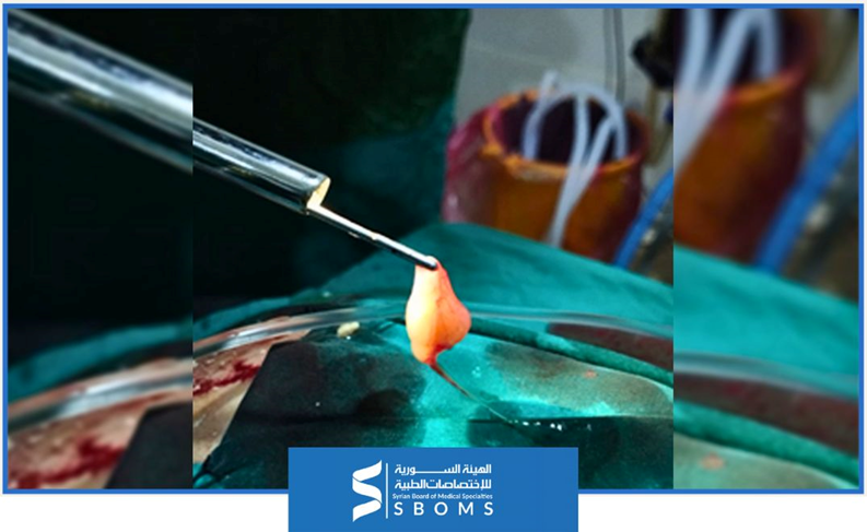
- A colloid cyst attached to the roof of the third ventricle was identified.
- The cyst was initially decompressed to reduce its size, followed by complete removal using delicate surgical instruments.
Cerebrospinal fluid was released under high pressure, confirming the diagnosis of obstruction caused by the cyst.

- Follow-up in the intensive care unit:
After the surgery, the patient was transferred to the intensive care unit to monitor her condition thoroughly.
- Recommendations for Post-Discharge Follow-up:
- Periodic Follow-up: Regular check-ups at the neurosurgery clinic to monitor for any recurrence of the cyst or the development of new complications.
- Health Education: The patient and her family were instructed on recognizing critical neurological symptoms, such as severe headaches or limb weakness, that may require immediate medical attention.
- Wound Care: Maintain proper wound hygiene by keeping it clean and dry and follow the doctor’s guidelines for dressing changes to prevent infection.
Conclusion:
This case highlights the crucial importance of timely surgical intervention in treating colloid cysts that block cerebrospinal fluid flow, leading to serious complications such as hydrocephalus and neurological decline. With coordinated care and effective surgical treatment, favorable outcomes can be achieved, enhancing the chances of complete recovery.
Colloid cysts
Colloid cysts are rare, benign tumors typically found in the anterior superior portion of the third ventricle, near the foramen of Monro [2]. These cysts constitute about 1% to 2% of all intracranial tumors, with an estimated annual incidence of 3.2 cases per million people.[3]
Clinical symptoms
While many colloid cysts are asymptomatic, they can cause obstructive hydrocephalus, leading to severe neurological decline or even death. Common symptoms include:
- Headaches
- Nausea and vomiting
- Visual disturbances
- Dizziness or ataxia
- Cognitive impairment
Symptomatic patients are typically younger, often between 30 and 40 years old. Hydrocephalus is a frequent and serious complication associated with these cysts.
Mechanisms of Neurological Deterioration:[4]
- Hydrocephalus: The colloid cyst can obstruct the cerebrospinal fluid (CSF) flow, increasing intracranial pressure, which may result in brain herniation.
- Cerebrospinal Fluid Obstruction: If the cyst detaches from its peduncle, it can completely block the foramen of Monro, leading to acute hydrocephalus and rapid neurological decline.
- Intracystic Hemorrhage: Bleeding within the cyst can cause severe expansion of the cyst, leading to ischemia in the anterior brain due to disrupted blood flow.
Management strategies
Management of colloid cysts typically involves surgical intervention, particularly when symptoms are present. Surgical options include:
- Endoscopic Surgery: A minimally invasive approach that allows for the removal of the cyst through small incisions using an endoscope.
- Microsurgical Resection: A more traditional open surgery that offers direct access to the cyst, allowing for complete removal.
In cases where the cyst is small and asymptomatic, careful monitoring may be appropriate. However, if symptoms develop or hydrocephalus is detected, surgery is usually recommended to prevent further complications.
Summary
Although colloid cysts account for a small percentage of brain tumors, their potential to cause severe complications necessitates thorough evaluation and management. Recognizing the clinical risks and implications is essential for ensuring effective treatment and optimizing patient outcomes.
Clarification:
The surgery was successfully performed at Bab Al-Hawa Hospital, supported by the Syrian American Medical Society (SAMS). The surgical team included:
- Dr. Jalal Al-Najjar: Specialist in Neurosurgery and Brain Tumor Surgery, and Chairman of the Scientific Council of Neurosurgery.
- Dr. Kamal Al-Hamdo: Specialist Neurosurgeon.
- Dr. Ahmad Al-Daaboul: Neurosurgery resident at the Syrian Board of Medical Specializations (SBOMS).
References:
[1] A. Roberts, A. Jackson, S. Bangar, and M. Moussa, “Colloid cyst of the third ventricle,” JACEP Open, vol. 2, no. 4, 2021, doi: 10.1002/emp2.12503.
[2] J. G. Malcolm, “Book Review: Youmans and Winn Neurological Surgery, Eighth Edition,” Neurosurgery, vol. 91, no. 3, 2022, doi: 10.1227/neu.0000000000002077.
[3] T. L. Beaumont, D. D. Limbrick, B. Patel, M. R. Chicoine, K. M. Rich, and R. G. Dacey, “Surgical management of colloid cysts of the third ventricle: a single-institution comparison of endoscopic and microsurgical resection,” J Neurosurg, vol. 137, no. 4, 2022, doi: 10.3171/2021.11.JNS211317.
[4] T. L. Beaumont, D. D. Limbrick, K. M. Rich, F. J. Wippold, and R. G. Dacey, “Natural history of colloid cysts of the third ventricle,” J Neurosurg, vol. 125, no. 6, 2016, doi: 10.3171/2015.11.JNS151396.

